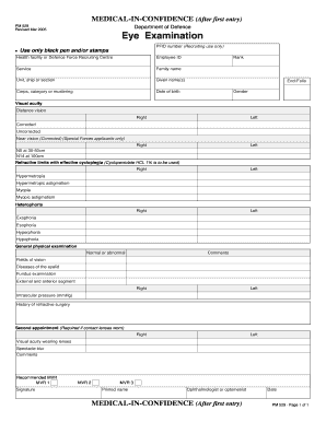
During this exam, the physician places drops in the eyes to make the pupils dilate (open widely) to allow a better view of the inside of the eye, especially the retinal tissue. figure 1. oct of a patient with bilateral proliferative diabetic retinopathy with diabetic macular edema. Aug 10, 2007 ocular examination: visual acuity: right eye (od) -not tested; left eye (os) -20/20; extraocular motility: full, . Then, assuming this baseline exam shows no pathology, dr. gerson generally dilates at intervals of retinal exam documentation every few years in young, healthy patients who have no family history of retinal disease. for viewing the retina, newer generation non-contact slit lamp lenses are gaining increased popularity. Shop vision screening & diagnostics from hillrom. we are a leading provider of medical technologies for the health care industry.

Uc San Diegos Practical Guide To Clinical Medicine
Oct 31, 2017 screening or monitoring for diabetic retinal disease refers to a retinal or dilated eye exam performed by an eye care professional (optometrist or . Since the mid-19th century, it has been recognized that changes in the optic nerve appearance retinal exam documentation correlate with vision and visual field loss in glaucoma. although there have been variations in the definition of glaucoma over time, increased attention to the structure and appearance of the optic nerve has been a hallmark in understanding glaucoma. May 24, 2016 the key to any examination is to be systematic and always perform each element. 1. visual acuity. in the clinic, visual acuity is typically .
• documentation of a negative retinal or dilated eye exam by an eye care professional in the year prior, where results indicate retinopathy was not present. documentation does not have to state specifically "no diabetic retinopathy" to be considered. Oct 30, 2020 · a retinal artery occlusion (rao) is a blockage in one or more of the arteries of your retina. the blockage is caused by a clot or occlusion in an artery, or a build-up of cholesterol in an artery. this is similar to a stroke. there are two types of raos: branch retinal artery occlusion (brao) blocks the small arteries in your retina. Retinal exam assessment of visual acuity: the first part of the eye exam is an assessment of acuity. this can be done with either a standard snellen hanging wall chart read with the patient standing at a distance of 20 feet or a specially designed pocket card (held at 14 inches). Aug 24, 2020 · the dye travels through your blood vessels. a special camera takes photos of your retina as the dye travels throughout the vessels. this test shows if the retinal vein is blocked. people under the age of 40 with central retinal vein occlusion (crvo) may be tested to look for a problem with their blood clotting or thickening. how is crvo treated?.
Ccs Exam Prep Exam 1 2019 Flashcards Quizlet

Examination Of The Eyes And Vision Osce Guide Geeky Medics
Fundoscopic / ophthalmoscopic exam visualization of the retina can provide lots of information about a medical diagnosis. these diagnoses include high blood pressure, diabetes, increased pressure in the brain and infections like endocarditis. introduction to the fundoscopic / ophthalmoscopic exam. Documentation in the medical record must include both: 1. a note indicating the date when the hrhpv test was performed retinal eye exam performed: a retinal or. Causes. in general, retinal detachments can be retinal exam documentation categorized based on the cause of the detachment: rhegmatogenous, tractional, or exudative. rhegmatogenous (reg ma todge uh nus) retinal detachments are the most common type. they are caused by a hole or tear in the retina that allows fluid to pass through and collect underneath the retina, detaching it from its underlying blood supply.
We can correctly presume that the most commonly used new patient code in ophthalmology is a comprehensive eye exam (92004). this is considered an easily documented code and the requirements fit the usual work of a retinal exam. the rules are straightforward: one must document a chief complaint, a medical history relevant to the reason for visit. Retinal hemorraghes: • what type of testing is useful to show retinal hemorraghing? • did you conduct this test? • what conclusions, if any, did you draw from the findings of this test? • what is the difference between unilateral and bilateral retinal hemorraghes? subdural hematoma:. Fundal examination should be an integral part of any eye examination. the cup/ disk ratio is slightly larger in the african american population. the normal fundus .
Vision Screening Diagnostics Hillrom
In the context of office visit documentation, the dilated posterior segment examination must be documented before a physician can bill separately for extended . And unlike the exam, no machine can “give a sense of comfort and satisfaction to the patient. ” but these sophisticated systems have enhanced clinical practice. “our understanding of retinal and macular disease is much more clearly defined,” dr. sarraf said. and there’s more to discover. “there’s always mystery involved in the. Sep 08, 2020 · with branch retinal vein occlusion (brvo), vision usually worsens due to swelling of the macula. the main goal of treatment is to dry up the retina. in most cases, medication or laser help reduce fluid and swelling. your ophthalmologist may also choose to treat your brvo with medication injections in the eye. the medicine can help reduce the swelling of the macula. Retinal exam; assessment of visual acuity: the first part of the eye exam is an assessment of acuity. this can be done with either a standard snellen hanging wall chart read with the patient standing at a distance of 20 feet or a specially designed pocket card (held at 14 inches). each eye is tested independently (i. e. one is covered while the.
Jan 7, 2019 2022f dilated eye exam with interpretation by an optometrist or ophthalmologist documented and reviewed; 2024f seven (7) standard field . Documentation in the record reveals that a patient is admitted with an acute exacerbation of copd (ms-drg 192). a higher-paying drg may be appropriate if documentation is present in the record at the time the decision was made to admit the patient that confirms a diagnosis associated with which of. Eye exam results: foveal retinal lesion(s) diagnosis/cause: perifoveal retinal burn and/or hemorrhage [bleeding]. lps commentary: this is more serious because the spot is at or close to the center of vision. the fovea is the central 2 percent of vision, about the width of a word or two read at arm’s length. This assessment is part of the nursing head-to-toeassessment you have to perform in nursing the eye assessment includes: inspection of the eyes for abnormalities, testing the if all these findings are normal you can document pe.
2019 hedis reference guide for primary care.
Comprehensive Hedis1 Documentation And Coding Guide

Documentation requirements to meet the measure:it is best if the pcp has a copy of the retinal eye exam in the patient record. you can document the year and results of the eye exam in your notes based on patient information, however this will not be picked up electronically for reporting. 6. external examination. look for any ptosis by measuring the margin-to-reflex distance, which is the distance from the corneal light reflex to the margin of the upper lid. look for lagophthalmos. note any unusual growths or lesions that may require a biopsy. palpate lymph nodes and the temporal artery if indicated by the history or exam.
Fluorescein angiography may be used to check for leaks, blocks, and abnormal growth retinal exam documentation of blood vessels. another test, optical coherence tomography (oct) provides detailed images of the thickness of the retina and allows the physician to measure swelling. vision tests may also be conducted. To help organize your eye exam, i've made a sample ophthalmology note on the facing page. in our notes we typically comment on four retinal findings:. Mar 1, 2020 once you are sure the patient truly meets “new” status, a properly documented patient retinal exam will often meet the requirements of 3 codes .
Diabetic retinopathy the american society of retina.
0 Response to "Retinal Exam Documentation"
Post a Comment